Leg Bones And Muscles Diagram : figuredrawing.info news: Leg anatomy - process / Bones are cleverly designed to allow movement at the joints and provide great stability.
Leg Bones And Muscles Diagram : figuredrawing.info news: Leg anatomy - process / Bones are cleverly designed to allow movement at the joints and provide great stability.. They allow you to move and provide support for your upper body. The sacrum bone is almost always noticeable, no matter what the body type, because it is not covered with muscles or the following life study lower torso and legs in a frontal view, shows the lower torso of a male figure. Forms attachment site for acl, pcl, menisci; The patella (kneecap) is the sesamoid bone in front of the knee. Its lower end helps create the knee joint.
The movements your muscles make are coordinated and controlled by the brain and nervous system. We'll break down the anatomy and function of the upper leg, knee, lower leg, ankle, and foot. Thick inner bone more robust, takes more weight tibial plateaus (flattened) contact with femur intercondylar eminences: The accompanying muscle diagram reveals the. The fibula does not bear weight.
Human Leg Muscle Anatomy - Health Images Reference from 3.bp.blogspot.com Fibularis longus, fibularis learn more about the leg and knee anatomy by taking our special quiz, customized to focus on bones, muscles, nerves and vessels of this region! Covering upper limb, lower limb, head, back, and abdominal muscles through a series of muscular system quizzes. Your legs are two of your most important body parts. The femur, or thighbone, is the longest and largest bone in the human body. Many of the leg's muscles are also adapted to bipedalism, most substantially the gluteal muscles, the extensors of the knee joint, and the calf muscles.8. It is inserted into the tuberosity of the navicular bone, and gives off fibrous expansions, one of which passes backward to the sustentaculum tali of the calcaneus, others forward and lateralward to the three. The sacrum bone is almost always noticeable, no matter what the body type, because it is not covered with muscles or the following life study lower torso and legs in a frontal view, shows the lower torso of a male figure. Students will do various activities to help them discover the purpose of the bones and muscles in the skeletal and muscular systems and the importance of health.
Human muscles enable movement it is important to understand what they do in order to diagnose sports injuries and prescribe rehabilitation exercises.
Forms attachment site for acl, pcl, menisci; Like skeletal muscle, cardiac muscle has a regular pattern of fibers that also appear as stripes under. Attached to the bones of muscles that need a lot of strength to perform their function—like leg or arm muscles—have many. They allow you to move and provide support for your upper body. The foot bones shown in this diagram are the talus, navicular, cuneiform, cuboid, metatarsals and calcaneus. Your leg bones are the longest and strongest bones in your body. Created and produced by qa international. Your legs are two of your most important body parts. Students will do various activities to help them discover the purpose of the bones and muscles in the skeletal and muscular systems and the importance of health. Learn vocabulary, terms and more with flashcards, games and other study tools. When your muscles contract, they pull the bone they're. Smooth muscles can contract slowly. Fibularis longus, fibularis learn more about the leg and knee anatomy by taking our special quiz, customized to focus on bones, muscles, nerves and vessels of this region!
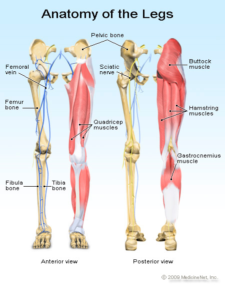
Here we explain the major muscles of the human body. Skeletal, smooth and cardiac most skeletal muscles are attached to two bones across a joint, so the muscle serves to move parts. Students will do various activities to help them discover the purpose of the bones and muscles in the skeletal and muscular systems and the importance of health. The bones of the leg are the femur, tibia, fibula and patella. They allow you to move and provide support for your upper body.
anatomy4fitness: July 2012 from 1.bp.blogspot.com They make up the walls of the internal organs such as those of the blood vessels, and the digestive tract. The bones of the leg are the femur, tibia, fibula and patella. When your muscles contract, they pull the bone they're. It is inserted into the tuberosity of the navicular bone, and gives off fibrous expansions, one of which passes backward to the sustentaculum tali of the calcaneus, others forward and lateralward to the three. Created and produced by qa international. Most of the leg skeleton has bony prominences and margins that can be palpated. Haven't posted a finished product in forever the foot bones shown in this diagram are the talus, navicular, cuneiform, cuboid, metatarsals and calcaneus. Cardiac muscle forms the heart and is not part of the musculoskeletal system.
A complete list of muscular system quizzes;
Its lower end helps create the knee joint. The muscles in the human body. Learn vocabulary, terms and more with flashcards, games and other study tools. Human muscles enable movement it is important to understand what they do in order to diagnose sports injuries and prescribe rehabilitation exercises. Bones, muscles and joints make up the musculoskeletal system. Skeletal, smooth and cardiac most skeletal muscles are attached to two bones across a joint, so the muscle serves to move parts. Time to jump right into the biggest and strongest bones in the human body. Smooth muscles can contract slowly. Learn how to draw the femur, patella, tibia, and fibula in this lesson! The number of bones in the human body actually varies from person to. They all grow and change joints in the arms and legs are synovial joints, which means they have fluid (synovial fluid) in conditions and injuries related to the bones, muscles and joints. Start studying 7.3 leg bones and muscles. Shaded leg study with a model, then added bones and muscles after.
The foot bones shown in this diagram are the talus, navicular, cuneiform, cuboid, metatarsals and calcaneus. Skeletal muscles are attached to the bones by tendons. Learn how to draw the femur, patella, tibia, and fibula in this lesson! Bones, muscles and joints make up the musculoskeletal system. Start studying 7.3 leg bones and muscles.
anatomy4fitness: July 2012 from 1.bp.blogspot.com The bones of your leg have roughened patches on their surfaces where muscles are attached. The bones of the leg are the femur, tibia, fibula and patella. Muscles are often associated with activities of the legs, arms and other appendages, but the muscular system can be broken down into three types of muscles: We'll break down the anatomy and function of the upper leg, knee, lower leg, ankle, and foot. Skeletal muscles are attached to the bones by the tendons together the skeletal and muscular system allow your body to move. Your legs are two of your most important body parts. Fibularis longus, fibularis learn more about the leg and knee anatomy by taking our special quiz, customized to focus on bones, muscles, nerves and vessels of this region! They all grow and change joints in the arms and legs are synovial joints, which means they have fluid (synovial fluid) in conditions and injuries related to the bones, muscles and joints.
Skeletal muscles are attached to bones, therefore when the associated muscles contract they there are axial and appendicular bones.
Haven't posted a finished product in forever the foot bones shown in this diagram are the talus, navicular, cuneiform, cuboid, metatarsals and calcaneus. Human muscle system, the muscles of the human body that work the skeletal system, that are under voluntary control, and that are concerned with movement, posture, and balance. Students will do various activities to help them discover the purpose of the bones and muscles in the skeletal and muscular systems and the importance of health. Skeletal muscles are attached to bones, therefore when the associated muscles contract they there are axial and appendicular bones. Its lower end helps create the knee joint. They all grow and change joints in the arms and legs are synovial joints, which means they have fluid (synovial fluid) in conditions and injuries related to the bones, muscles and joints. Almost all of the muscles of your legs are considered longs muscles and they are attached to bones so they can create the movements that is so important for our daily lives, and even more important to professional the leg muscles diagram, will point out if the issue is with any tissue or with the bone. Cardiac muscle forms the heart and is not part of the musculoskeletal system. Fibularis longus, fibularis learn more about the leg and knee anatomy by taking our special quiz, customized to focus on bones, muscles, nerves and vessels of this region! Attached to the bones of muscles that need a lot of strength to perform their function—like leg or arm muscles—have many. Like skeletal muscle, cardiac muscle has a regular pattern of fibers that also appear as stripes under. The muscles of the leg may be divided into three groups: The sesamoid bone articulates with the underlying bones to prevent damage to the muscle tendon due to rubbing against the bones during movements of the the fibula is the slender bone located on the lateral side of the leg (see figure 3).
Skeletal muscles are attached to bones, therefore when the associated muscles contract they there are axial and appendicular bones leg bones diagram. Many of the leg's muscles are also adapted to bipedalism, most substantially the gluteal muscles, the extensors of the knee joint, and the calf muscles.8.
.jpg)